


ACS Nano | Mechanistic Study of the Biological Response of Polymer Nanoparticles
Polymeric nanoparticles show great potential for diagnostic integration and nanopharmaceutical applications. However, these nanoparticles often fail to fulfill their designed functions due to factors such as nonspecific biomolecule adsorption, nonspecific cellular uptake, and excessively short in vivo circulation times. Over the years, researchers have overcome these obstacles and improved the performance of polymer nanoparticles by controlling the physicochemical parameters of the nanoparticles (including size, shape, morphology, surface charge, hydrophilicity, elasticity, surface chemistry, etc.). However, to date, there have been few studies of nanoparticle bioresponsiveness based on a nanoparticle system simultaneously at the molecular, cellular, and organismal levels.
Recently Bin Liu's group at the National University of Singapore published its mechanistic study on the biological response of polymer nanoparticles in ACS Nano. The group characterized the interactions and biological behaviors of polymer nanoparticles with cytoplasmic proteins, epithelial cells and zebrafish. Based on the results, the group proposes a rational framework to develop polymer nanoparticles with optimized properties to simultaneously achieve desired functions at the molecular, cellular and organismal levels.
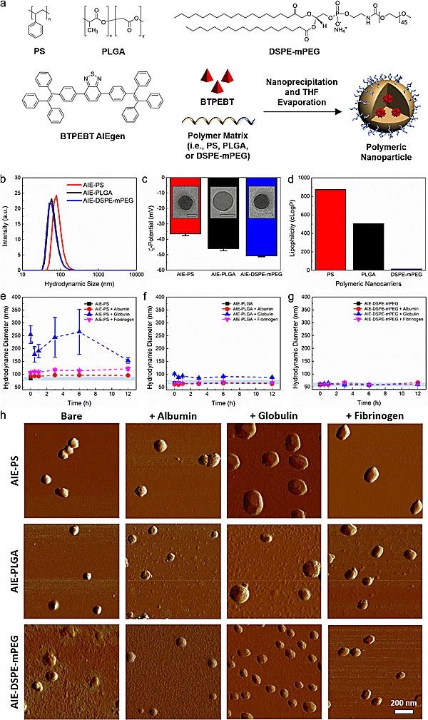
Figure 1. characterization of the interaction of different polymeric nanoparticles encapsulating AIE molecules with three major plasma proteins
The group used three different polymers (polystyrene PS, polylactic acid-hydroxyacetic acid PLGA, and lipid PEG derivative DSPE-mPEG), all of which were supplied by the above polymers from Chongqing Yu Yixing Pharmaceutical Technology Co. Coated with the fluorescent AIE molecule BTPEBT, three types of nanoparticles with diameters of about 60 - 80 nm were prepared and the interactions of the nanoparticles with the three major plasma proteins (albumin, globulin and fibrinogen) were investigated (Figure 1). The results showed that with the increase of time, the nanoparticles of AIE-PS and AIE-PLGA produced stronger interactions with plasma proteins with a several-fold increase in their hydrodynamic diameters, whereas no change in the diameter of the DSPE-mPEG nanoparticles was observed, which suggests that the DSPE-PEG possesses the weakest nanoparticle-protein interactions when it acts as a carrier for nanoparticles (Fig. 2).
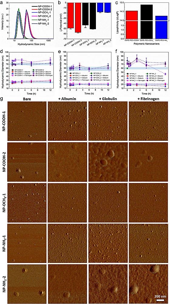
Fig. 2. Characterization of the interaction of AIE-DSPE-PEG nanoparticles with different surface functional groups with three plasma proteins
After discovering the inertness of AIE-DSPE-mPEG to plasma proteins, the group prepared a total of six nanoparticles with two sizes (20 nm NP-1, 60 nm NP-2) and three surface functional groups (carboxy-COOH, methoxy-OCH3, amino-NH2) (Fig. 3). As a result of the characterization, for the 60 nm nanoparticles, only globular proteins induced a slight increase in the size of NP-COOH-2; NP-OCH3-2 was unchanged by the interaction with all three proteins; and NP-NH2-2 showed a large size increase by the interaction with all three proteins. Interestingly, none of the NP-1 sizes change over time in the presence of plasma proteins, suggesting that plasma proteins are not adsorbed on the surface of the 20 nm polymer nanocarriers.
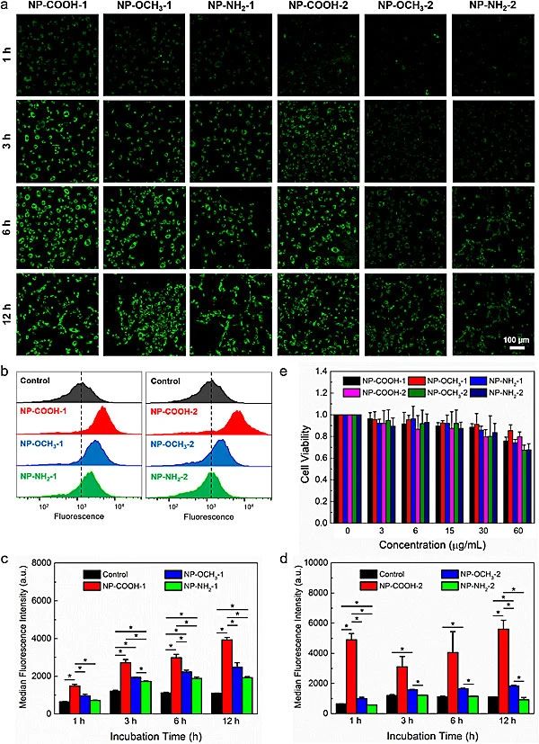
Figure 3.Characterization of cellular uptake and cytotoxicity of different nanoparticles in human umbilical vein endothelial cells (HUVEC)
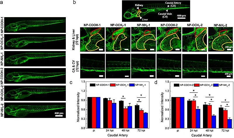
Figure 4. in vivo circulation and biodistribution of polymer nanoparticles in zebrafish
The group then investigated the cellular uptake and cytotoxicity of nanoparticles with human umbilical vein endothelial cells (HUVECs). Confocal microscopy imaging and flow cytometry results showed that HUVECs were more likely to take up smaller-sized nanoparticles. Comparing the three surface functional groups, the nanoparticles of NP-COOH with zeta potential zui low were easily taken up by the cells, on the contrary NP-NH2 with higher zeta potential was more difficult to be taken up by the cells. All nanoparticles showed good biocompatibility. The group also investigated the in vivo cycling and biodistribution of different nanoparticles in zebrafish model (Figure 4). The results showed that the circulation time and biodistribution of the polymer nanoparticles depended on their physical size. Smaller nanoparticles possessed longer in vivo circulation time. For a given nanoparticle size, the most negatively charged nanoparticles modified with COOH functional groups showed the longest circulation time, while the least negatively charged NP-NH2 had the shortest circulation time.
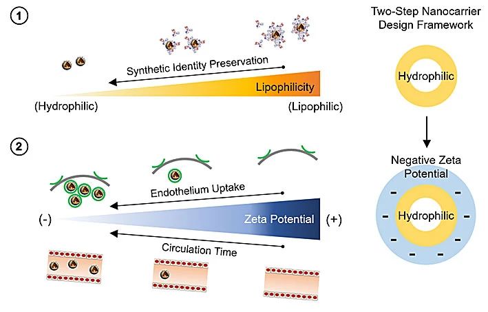
Fig. 5. Schematic framework of two-step nanocarrier design utilizing lipophilicity and zeta potentials
Based on the above studies, the group proposed a two-step design framework (Fig. 5) for the preparation of polymer nanoparticles with desired in vitro and in vivo biological behaviors. First, the nanocarriers should be designed to be as hydrophilic as possible to minimize nonspecific biomolecule adsorption and maintain their properties. Next, hydrophilic nanocarriers should be provided with a more negative zeta potential to enhance their cellular uptake and in vivo circulation time for better imaging and therapeutic effects.
Note: This article is the editor of this site reproduced, reproduced for the purpose of transmitting more information, and does not mean that we agree with its views and is responsible for its authenticity. If it involves the content of the work, copyright and other issues, please contact this site within 30 days, we will remove the content at the first time! [Statement] This site article copyright belongs to the original author of all content for the author's personal views This site only provides reference and does not constitute any investment and application recommendations. This site has the final interpretation of this statement